Figures & data
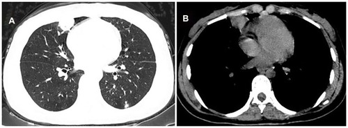
Figure 1 Case 1. CT findings in the right-superior pulmonary lobe. (A and B) A 1.7×1.4cm nodule was found at the initial diagnosis of pulmonary tuberculosis. (C and D) The nodule was almost absorbed after six months of chemotherapy.
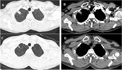
Figure 2 Case 1. Time course of CT findings in the right-inferior pulmonary lobe. (A and B) There was no obvious mass at the initial diagnosis of pulmonary tuberculosis. (C and D) A new occupying lesion in diameter of 3.4cm×2.3cm appeared after six months of chemotherapy, and its margin was irregular. (E and F) The mass was diminished to 1.5cm×1.3cm seven months after the initial diagnosis. (G and H) The mass was 0.9cm×1.1cm nine months after the initial diagnosis.
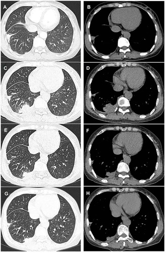
Figure 3 Case 1. CT-guided percutaneous lung biopsy shows caseous necrosis and granulomatosis surrounded by epithelioid and multinucleated giant cells. Hematoxylin and eosin, ×400.
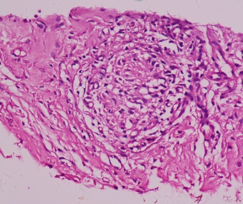
Figure 4 Case 2. CT findings in the right middle and inferior pulmonary lobe. (A and B) A 3.9cm×2.9cm lobulated mass without pleural effusion was found was after four months of anti-tuberculosis treatment.
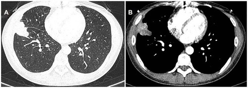
Figure 5 Case 2. CT-guided percutaneous lung biopsy shows plentiful infiltration of epithelioid cells and lymphocytes. Hematoxylin and eosin, ×400.

Figure 6 Case 3. Time course of CT findings in the left-superior pulmonary lobe. (A and B) A new occupying lesion in diameter of 1.4cm×0.8cm appeared after six months of chemotherapy with irregular margin and central low density. (C and D) The nodule was diminished to 1.0cm×0.3cm eight months after the initial diagnosis.
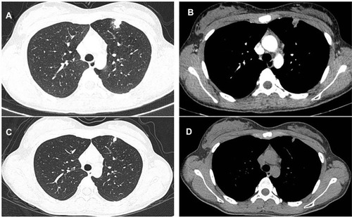
Figure 7 Case 4. CT findings in the right-inferior pulmonary lobe. (A and B) A mass measuring 3.3cm×2.0cm was found after three months of anti-tuberculosis therapy. (C and D) The mass was irregular with a cavity.

Table 1 The Clinical Characteristics Of Five Patients
Figure 8 Case 5. CT findings in the right middle pulmonary lobe. (A and B) A 2.0cm×1.5cm irregular mass appeared after two months of anti-tuberculosis treatment.