Figures & data
Table 1 Complete Blood Count
Table 2 Coagulation Studies And Myocardial Markers
Table 3 Serum Biochemistry Levels
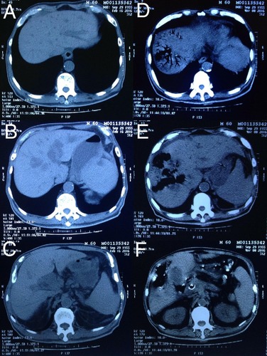
Figure 1 (A)–(C) Abdominal computed tomography (CT) before stenting revealed intrahepatic duct dilation due to obstruction of pancreatic mass. (D)–(F) Emergency CT showed gas-containing lesions in the liver, containing liver parenchymal moth-eaten destruct and pneumatized bile duct without obvious inflammation around. (F) The stent was right in the common bile duct without obstruction.