Figures & data
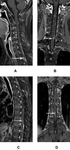
Figure 1 The irregularly thickened meninges of intraspinal tuberculosis show obvious homogeneous enhancement on MR images.
Notes: Intraspinal tuberculosis in a 21-year-old male. Sagittal T2WI (A) shows a slight hyperintensity of the irregularly thickened meninges (white arrow) and edema of the involved spinal cord. Gadolinium contrast MR images (B–F) show obvious enhancement of irregularly thickened meninges in the thoracic and lumbar segments (white arrow) that integrate with each other. Nerve roots are thickened with obvious enhancement bilaterally (E, white arrow).
Abbreviations: T2WI, T2-weighted imaging; MR, magnetic resonance.
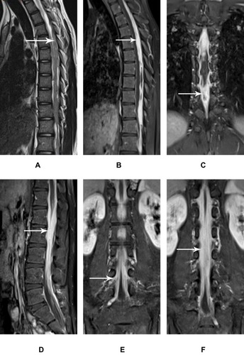
Figure 2 Multiple intraspinal tuberculomas companied with thickened meninges in the cervical, thoracic, and lumbar segment.
Notes: Intraspinal extramedullary tuberculomas in a 25–year–old female. Sagittal T2WI (A, D) shows a slight hyperintensity of the tuberculomas (white arrow) and diffuse edema of the spinal cord. Gadolinium contrast MR images (B–C, E–F) show tuberculomas with both homogeneous (C, white arrow) and ring (E, white arrow) enhancement that integrate with adjacent irregularly thickened meninges. Cervical lymph nodes are enlarged and exhibit ring enhanced (C, white arrowhead).
Abbreviations: T2WI, T2-weighted imaging; MR, magnetic resonance.
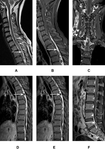
Figure 3 Multiple intraspinal metastatic lesions accompanied with slightly irregularly thickened meninges.
Notes: Intraspinal metastases in a 60–year–old female with breast cancer. Sagittal T2WI (A) shows slightly hyperintense intramedullary nodularities in the thoracic region (white arrow) accompanied by edema in the adjacent spinal cord. Gadolinium contrast MR images (B–F) show multiple intraspinal subdural lesions which are closely adherent to the slightly thickened meninges (white arrow).
Abbreviations: T2WI, T2-weighted imaging; MR, magnetic resonance.
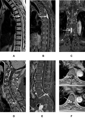
Figure 4 Multiple intraspinal metastatic lesions accompanied by patchy thickened meninges.
Notes: Intraspinal metastases in a 64–year–old female with breast cancer. Gadolinium contrast MR images (A–D) show obviously enhanced nodules closely adherent to the slightly thickened meninges; the lesions have sharp margins and clear distinction between them (white arrow).
Abbreviations: T2WI, T2-weighted imaging; MR, magnetic resonance.