
Figures & data
Figure 1 Morphology and Gram’s staining of P. sputorum. Culture blood samples were transferred to Columbia blood plates and incubated in the 5% CO2 incubator at 37°C for (A) 24 h and (B) 48 h. Gram’s staining showed that P. sputorum is (C) a gram-negative bacilli.
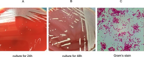
Figure 2 Phylogenetic analysis. Phylogenetic trees based on 16S rRNA gene sequences indicated the phylogenetic positions of isolates 1 and 2 and of other Pandoraea species. Burkholderia sordidicola S5-BT12826 was used as the outgroup. Bootstrap values (>70%) are shown for appropriate nodes. The scale bars represent the number of nucleotide substitutions per site.
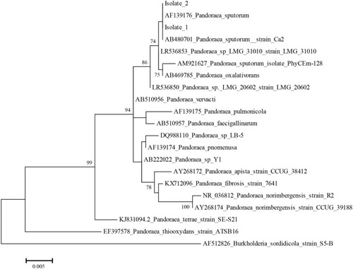
Figure 3 Relatedness of the two isolates as determined by pulsed-field gel electrophoresis. Chromosomal DNA of the two isolates was digested with restriction endonucleases xbal and spel, followed by pulsed-field gel electrophoresis. (A) Lane 1, Marker; Lane 2, Isolate 1; Lane 3, Isolate 2. (B) Lane 1, Marker; Lane 2, Isolate 1; Lane 3, Isolate 2.
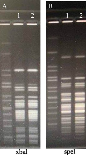
Table 1 Antibiotic Susceptibility Patterns Of P. Sputorum Isolates Described In This And Previous Studies