Figures & data
Figure 1 Antibiotic resistance pattern of all isolated P. aeruginosa isolates.
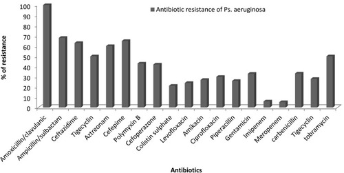
Figure 2 Phenotypic detection for mcr positive isolates by combined disc diffusion test (CDT). (A): mcr-1 positive strain showed an increase in the zone diameter of discs with colistin and EDTA ≥ 3mm in comparison to colistin alone. (B): mcr-1 negative isolate showed slight change (1 mm) in the inhibition zone diameter of colistin and EDTA disc in comparison to colistin alone.
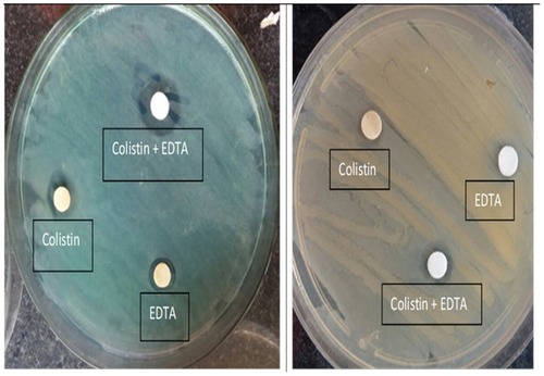
Figure 3 Antibiotic resistance pattern of colistin-resistant isolates.
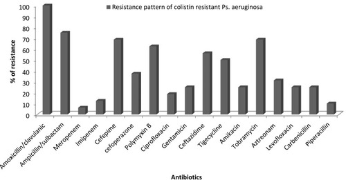
Table 1 Colistin-Resistant Isolates, Some Possible Mechanisms of Resistance to Colistin and Their Susceptibility to Other Antibiotics
Table 2 Molecular Weights and Amount % of Extracted Outer Membrane Proteins of Colistin Resistant and Colistin Sensitive P. Aeruginosa
Figure 4 Outer membrane SDS-PAGE of colistin resistant and sensitive strains. Lane 1: Protein Marker, Lane 2 and Lane 3: colistin-resistant strains (P1 & P12), Lanes 4–6: colistin sensitive strains.
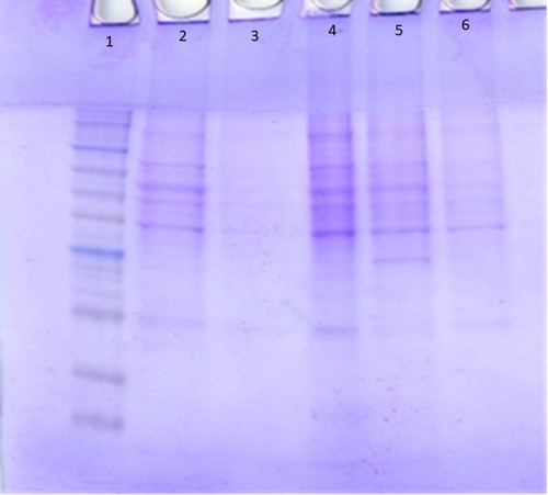
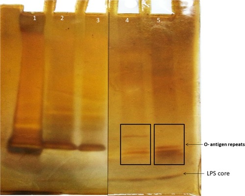
Figure 5 LPS bands pattern. Lanes 1, 2 & 3: colistin-resistant mcr-1 negative strains (P3, P6 & P10, respectively), Lane 4: Colistin sensitive strains and Lane 5: Colistin-resistant mcr-1 positive strain (P1). O-antigen repeats are boxed and arrow refers to LPS core.