Figures & data
Figure 1 The latex drainage tubes were inserted into the wound. Note that the red lines indicate the direction and depth of the latex side-holed drainage tubes, some of which were pulled through the dissected pathway to form a through-and-through drain.
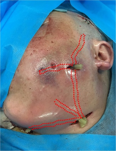
Figure 2 The foam materials were cut into bar-shaped pieces and inserted into the wound along with the latex drainage tubes. Note that the blue lines indicate the direction and depth of the foam.
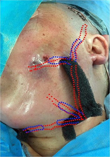
Figure 3 The bar-shaped foam materials were connected by a block of foam dressing on the surface of the face and neck. Sutures were sometimes needed to stabilize the foam block.
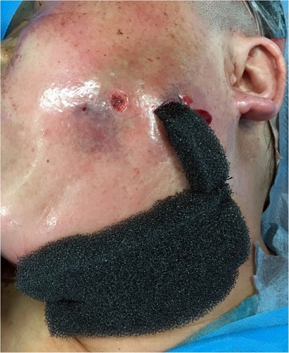
Figure 4 After the application of the film, an adhesive disc and drainage tube were connected to the foam dressing with a pressure-controlled vacuum source.
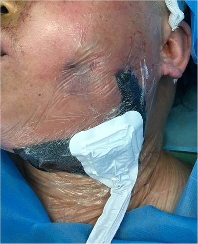
Figure 5 The drainage was efficient and the container of the NPWT device needed to be replaced once it was almost full.
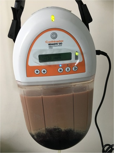
Table 1 The Demographics and Clinical Characteristics of the Patients
Figure 6 This patient recovered 4 days after the removal of the NPWT device, leaving the scars to be corrected later.
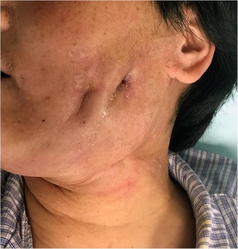
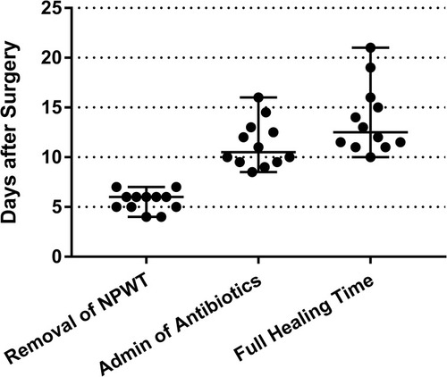
Figure 7 Scatter plot of the time of removal of NPWT, administration of antibiotics, and complete wound healing of all 12 patients. The median time and range are indicated.