
Figures & data
Figure 1 (A, B) 18F-fluorodeoxyglucose positron emission tomography/computed tomography (18F-FDG-PET/CT) detected the presence of an FDG-avid tumor (arrow) in the ascending colon (A) and peritoneal nodules (arrowheads) (B). (C) A biopsied peritoneal nodule showed epithelioid cell granuloma with partial necrosis on hematoxylin-eosin staining. (D) Abdominal CT showed a thickened peritoneum with massive ascites at the onset of tuberculosis peritonitis.
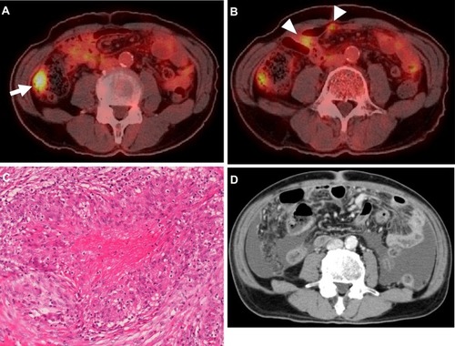
Table 1 List of Reported MPN Patients Complicated by Mycobacterium Tuberculosis (MTB) Infection During Treatment with Ruxolitinib