
Figures & data
Figure 1 Workflow for identification of underlying disease favoring invasive nocardiosis.Citation17
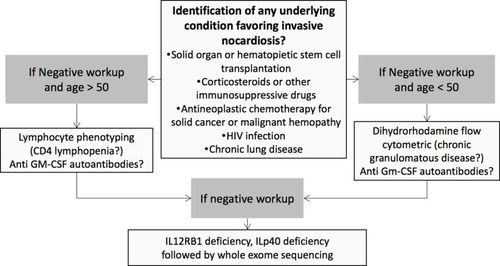
Figure 2 Radiographic findings in patients with invasive nocardiosis. (A) Chest CT-scan of a sixty-year-old patient with chronic lymphocytic leukemia treated with ibrutinib who developed Nocardia pneumonia. (B and C) Twenty-one-year-old patient with chronic granulomatous disease who developed Nocardia pulmonary abscess with local extension to the ribs (white arrow). (D) Brain MRI of a forty-six-year-old cardiac transplant patient who developed Nocardia brain cerebral abscess (white arrowhead): ring-enhancing multilobulated lesion surrounded by edema causing a mass effect on the anterior ventricles. MRI, axial T1 after gadolinium injection.
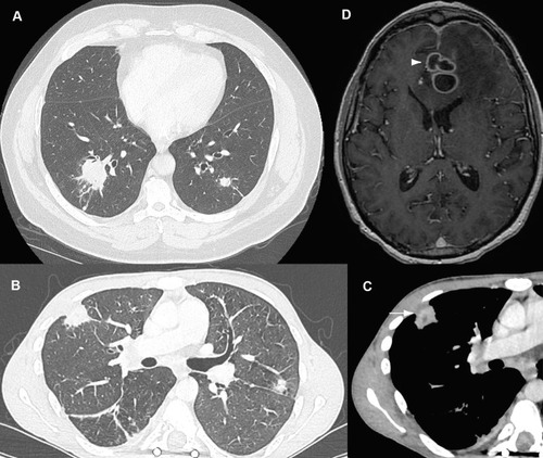
Figure 3 Microbiological diagnosis of nocardiosis. (A) Direct examination of a bronchoalveolar lavage (BAL) after Gram staining revealing filamentous Gram-positive bacteria (white arrowhead). (B) Positive culture of the same BAL on blood agar plate.
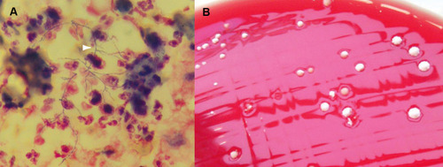
Table 1 Results of Antibiotic Susceptibility Testing Among Nocardia Isolates According to Their SpeciesCitation3,Citation48,Citation49,Citation67–Citation69
Table 2 Main Characteristics of Antibiotics That Can Be Used Before Obtaining the Results of Antibiotic Susceptibility Testing, ie, for the Initial Antibiotic TreatmentCitation2,Citation62,Citation70
Table 3 Proposed Initial Treatment and Antibiotic Duration for Invasive Nocardiosis, Based on Clinical Presentation, Before Obtaining Species Identification and/or Antibiotic Susceptibility Testing