Figures & data
Figure 1 Chest radiography and computed tomography images. (A, B, and C) were obtained six, four, and one month(s) before admission, respectively (black arrow indicates interstitial infiltration); (D1) multiple ground-glass opacities on admission; (D2–5) multiple nodular lesions with cavities on both lung fields on admission.
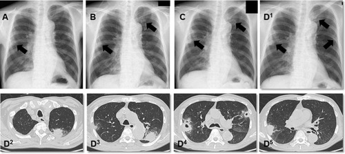
Figure 2 Gram-stain of bronchoalveolar lavage. Black arrows indicate capsulated Gram-negative large rod.
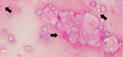
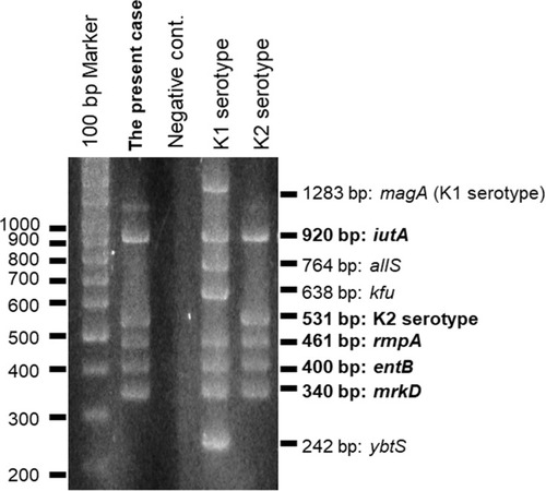
Figure 3 Multiplex PCR. The isolated strain possessed iutA, rmpA, entB, and mrkD.