Figures & data
Table 1 Primer Sequences Used for Amplification of (Nuc) and (mecA) Genes and the Suspected Product Size
Figure 1 (A) Amplified PCR products of nuc gene at (270 bp). Lane 1: 100 bp ladder, Lanes 2–4: positive to Staphylococcus aureus; (B) Amplified PCR products of mecA gene at (147 bp). Lane (M) 100 bp ladder, Lanes 1–6: positive to mecA gene.
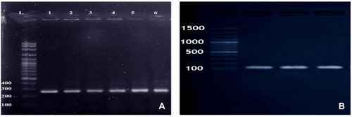
Table 2 Re-Isolated MRSA Strains from Internal Organs Following Oral Challenge of Mice
Figure 2 Histopathological changes in liver of challenged mice. (A) Liver showing patchy area of hepatic cellular necrosis mixed with polymorph nuclear and mononuclear inflammatory cells (arrow), portal congestion, bile duct hyperplasia and leukocytic aggregations mostly, with neutrophils, macrophages and lymphocytes (H&E X400). (B) Liver showing multiple focal areas of hepatocellular necrosis with neutrophils (arrows) and few mononuclear cell aggregation, sinusoidal dilation and kupffer cells activation (H&E X400). (C) Liver showing focal area of hepatic cellular necrosis (rectangle) mixed with polymorph nuclear, mononuclear inflammatory cells and fragmented nuclei (H&E X400). (D) Liver showing intense periportal inflammatory cell aggregation (arrow) (H&E X400). H&E, hematoxylin and eosin stain.
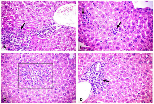
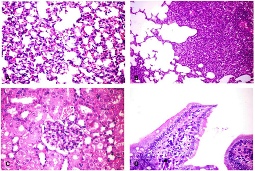
Figure 3 Histopathological changes in different organs of challenged mice. (A) Lung showing thickening of the alveolar wall by dilated perialveolar blood capillaries, inflammatory cells mainly neutrophils and mononuclear cells (H&E X400). (B) Lung showing severe fibrinopurulent pneumonia with giant alveoli (H&E X400). (C) Kidney showing glomerular hypertrophy with mesangial hypercellularity (H&E X400). (D) Intestine showing sloughing and desquamation of individual enterocytes with increased lamina propria macrophages, lymphocytes and neutrophils (H&E X200). H&E, hematoxylin and eosin stain.