
Figures & data
Figure 1 Radiological findings of the present case. Chest X-ray revealing lobar pneumonia on bilateral lung fields (A). Computed tomography showing multiple consolidation with air-bronchogram, nodules, and ground-glass attenuation on bilateral lung fields (B1–4).
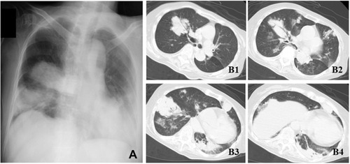
Figure 2 Multiplex PCR. The present strain harbored iutA, rmpA, entB, and mrkD genes.
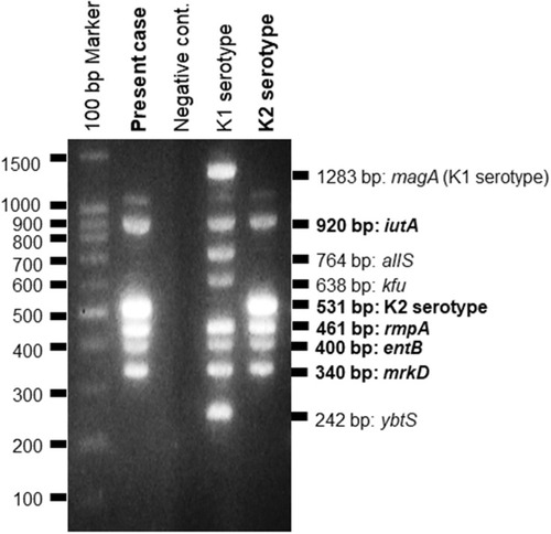
Table 1 The Characteristics of Nine Adult Cases with Community-Acquired Pneumonia Due to Hypervirulent Klebsiella pneumoniae Including the Present Case [14–16]