Figures & data
Table 1 Colony Diameters of Tryptophol (TOH)-Treated Scedosporium apiospermum CBS 117410 Following Incubation at 25°C
Figure 1 Culture of S. apiospermum CBS 117410 on Sabouraud Dextrose Agar (SDA), (A) front view, (B) reverse view, and (C) macroconidia of S. apiospermum stained by lactophenol cotton blue. Scale bar of A, B = 100 μm and C = 20 μm.
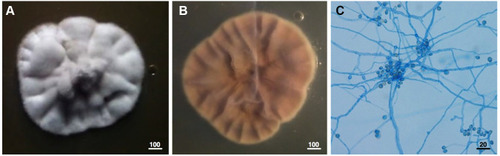
Figure 2 Coating catheter with Tryptophol (TOH) significantly reduced the intraluminal biofilm formation of Scedosporium apiospermum CBS 11741 (108 conidia/mL) after 48 h post-inoculation as observed under a scanning electron microscope (SEM). Scanning electron micrograph in the luminal surface of (A) uncoated catheter, (B) uncoated catheter with S. apiospermum, (C) voriconazole-coated-catheter (100 μM) with S. apiospermum, and (D) TOH-coated-catheter (100 μM) with S. apiospermum. Scale bar (A–D) = 20 μm.
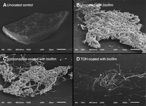
Figure 3 Biofilm of Scedosporium apiospermum CBS 11741 (108 conidia/mL) in the luminal surface of tryptophol (TOH)-coated catheter after 12 and 48 h post-inoculation as examined by (A) H-score, (B) crystal violet, and (C) XTT assays. Results are presented as mean ± standard deviation (SD). *,#Indicated as significant difference at p < 0.05 by 2-way ANOVA.
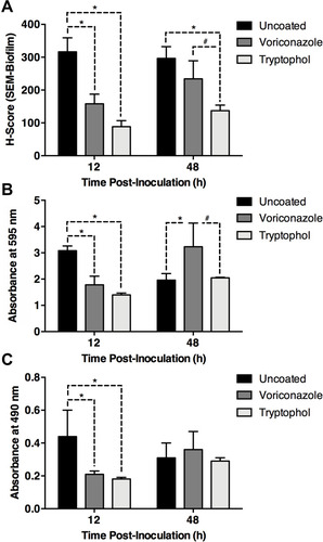
Figure 4 Brain histopathological studies in catheterization rats with Scedosporium apiospermum CBS 11741 infections after 48 h post-inoculation. In uncoated catheter groups, (A) brain abscess was developed without encapsulation. (B–E) The perivascular cuffing mainly with neutrophil infiltrates were found around the site of infection with the presence of S. apiospermum hyphae (as indicated by the arrows in B) and (F) in meninges. (G–I) Unlike the brain without abscess in tryptophol (TOH)-coated catheter groups, absent of necrotic area surrounded with inflammatory cells were noted compared to infected brains. Scale bar = 100, 20, and 10 μm.
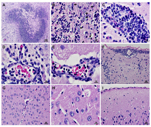
Figure 5 Lung histopathological studies in catheterization rats with Scedosporium apiospermum CBS 11741 infections after 48 h post-inoculation. In uncoated catheter groups, (A) severe hemorrhage (haemothorax) and (B) the presence of inflammatory cells including lymphocytes and macrophages were seen. In tryptophol (TOH)-coated catheter groups, (C and D) absent of severe hemorrhage and inflammatory cells were noted. Scale bar = 100 and 20 μm.
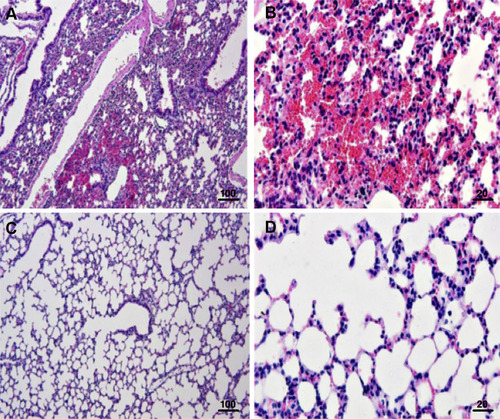
Table 2 Effect of S. apiospermum-Infected Rat Serum on the Survival of G. mellonella
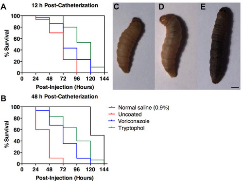
Figure 6 S. apiospermum infected rat sera from TOH-coated catheter extended G. mellonella lifespan. Data represented percentages of survival of G. mellonella larvae treated with S. apiospermum infected rat sera after (A) 12 h post-injection and (B) 48 h post-injection. A percentage of survival in each condition was also compared to normal saline (0.9%). (C) G. mellonella larvae showed melanized in 1–2 h post-injection. (D) Melanization of G. mellonella larvae cuticle was darker and (E) completely black near the time of death. Scare bar = 1 mm. Lifespan was analyzed using Kaplan–Meier analysis and p - values were calculated using Log-rank test. Statistical details of lifespan are summarized in .