Figures & data
Table 1 General Clinical Data of the 263 Included Cases
Figure 1 Imaging findings of chest wall tuberculosis (TB). (A) Sinus formation caused by TB lesions piercing the chest wall. (B) Chest wall TB lesion breached through the intercostal muscle to form a dumbbell-like abscess. (C) In some patients with chest wall TB, the lesion showed no signs of healing after surgical treatment (no anti-TB treatment). (D) Secretion at the incision site. (E) Chest wall TB lesion only formed in local thoracic masses. (F) After the patient depicted in panel E underwent chest wall lesion removal, his lesion showed no residue and the wound healed well.
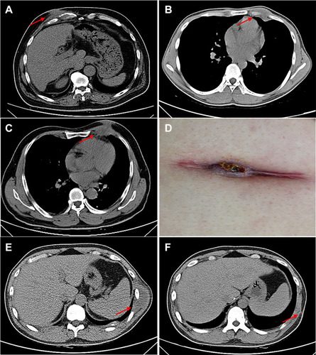
Table 2 Anti-Tuberculosis Time Comparison
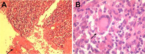
Figure 2 Histopathological sections of chest wall TB lesions, showing necrotic material (A) and granulomatous lesions with multinucleated giant cells (B). Pathology confirmed that the lesion was tuberculous granulomatous inflammation.