
Figures & data
Figure 1 The chest CT showed nodules and patches in the upper lobe of the left lung on admission (A); enlarged lesions in the upper lobe of both lungs and bilateral pleural effusions were observed on the 13th day after admission (B); bilateral infiltrates, interstitial infiltrates, alveolar infiltrates and bilateral pleural effusion were observed on the 20th day after admission (C).
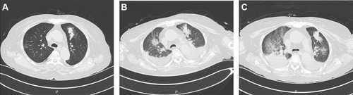
Figure 2 Yeast forms of Penicillium janthinellum in BAL washings stained with fluorescence (A, Original magnification X 400), Gram stains (B, Original magnification X 1000), hexamine silver (C, Original magnification X 400) and fungal morphology stained with medan lactate (D, Original magnification X 400). Colony morphology in the obverse side was cultured in 28 °C PDA medium for 5 days (E) and Colony morphology in the reverse side (F) was cultured in 28 °C PDA medium for 14 days.
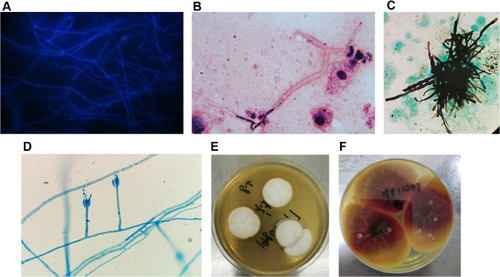
Table 1 Fungal Susceptibility