
Figures & data
Figure 1 Chest images. (A) Chest X-ray showing bilateral patchy, cloudy opacities in the lower lungs with some adjacent pleural thickening and adhesion before treatment. (B and C) Chest HRCT showing diffused stripes as well as reticular and ground-glass opacities with honeycomb structures, primarily in the lower lungs before treatment. (D and E) Chest HRCT scan showing a faint patchy opacity in the lateral segment (white arrow).
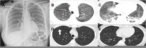
Table 1 Main Laboratory Test Results Before and After Treatment
Table 2 Reports of PCP Cases Detected by Metagenomics Next-Generation Sequencing (mNGS)