Figures & data
Table 1 Comparison of Clinical and Laboratory Information Between Patients with and without New Pulmonary Lesions
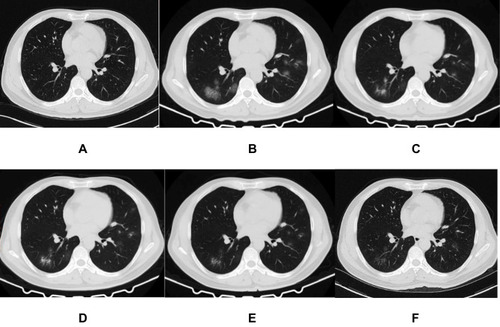
Figure 1 A 45-year-old woman who exposed to COVID-19 patients has cough and diarrhea for 2 days. The initial chest CT is negative (A). After 4 days, the first repeat CT shows a new patch GGO located in the left lower lobe with homogeneous density (B). Subsequently, this new lesion increases in extent and density (C), and then absorbs gradually (D and E). On the latest CT scan (31 days later), it significantly absorbs and leaves a small amount of faint GGO (F).

Figure 2 A 73-year-old man who exposed to COVID-19 patients has fever and fatigue for 2 days. The initial chest CT is negative (A). After 2 days, the first repeat CT shows multiple patchy GGOs with heterogeneous density developed in the bilateral lungs (B). Subsequently, some of new lesions increase in extent and density (C), and then absorbs gradually with decrease of extent and increase of density (D and E). On the latest CT scan (38 days later), they significantly absorb and leave few fibrous strips (F).
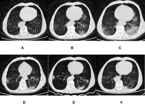
Figure 3 A 44-year-old man who exposed to COVID-19 patient has fever, cough, and diarrhea for 4 days. The initial chest CT is negative (A). After 7 days, the first repeat CT shows multiple spherical and patchy GGOs with heterogeneous density developed in the bilateral lungs (B). Subsequently, the lesions directly absorb with decrease of extent and temporary increase of density (C–E). On the latest CT scan (29 days later), they significantly absorb and leave a small amount of faint GGO (F).