Figures & data
Figure 1 Multiple separated hyperkeratotic nodular lesions with erythematic and prominent borders on the patient’s face.
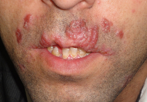
Figure 2 Histopathological assessment of lesions revealing skin tissue with multiple granuloma formation in the subepithelial area composed of epithelioid cells, Langhans giant cell, and lymphocyte around them (Magnifications: 10× and 40×).
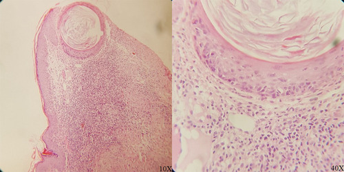
Figure 3 The Ziehl–Neelsen stained histopathological assessment (Magnifications: 10× and 40×).
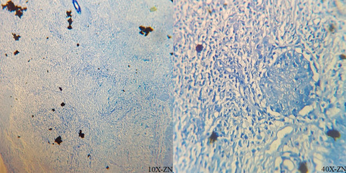
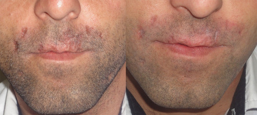
Figure 4 Clinical appearance of lesions at the end of the 6th (left) month of treatment, and after the second month after treatment (right).