
Figures & data
Figure 1 A chest computed tomography (CT) scan in Case 1 shows infiltrative and nodular shadows with pneumatocele formation in the right upper lobe.
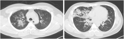
Figure 2 Bronchoscopy in Case 1 shows diffuse inflammatory changes and easy bleeding.
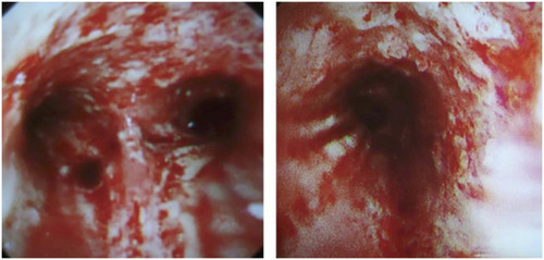
Figure 3 A chest CT scan in Case 2 shows irregular consolidations, ground glass opacity and bilateral pleural effusion.
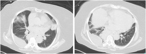
Table 1 Genetic Characteristics and Virulence Factors in Two Isolates in This Study