Figures & data
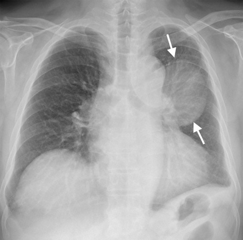
Figure 1 (A, B) The chest radiograph (A) and contrast-enhanced axial chest CT image with a mediastinal-window setting (B) obtained at our hospital showing a large mass (arrows in A and B) in the left upper lobe. The mass exhibits heterogeneous contrast enhancement with a central low-attenuation area. Note blunting of the left costophrenic angle secondary to left pleural thickening. There is no enlarged mediastinal or hilar lymph node.
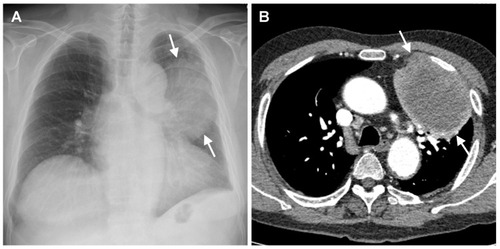
Figure 2 (A–D) Histopathology revealed (A) Tumor admixed with necrotic tissue (HE stain, 100x), (B) Gram-negative bipolar rods resembling safety pins (Brown-Hopp stain, 1000x), (C) Non-small cell carcinoma favor adenocarcinoma (HE stain, 200x), (D) TTF-1 immunohistochemistry positive nuclear staining (200x).
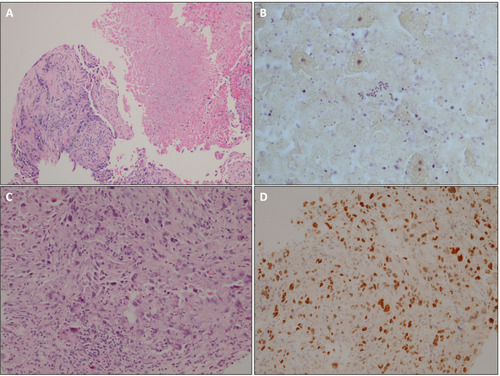
Figure 3 The follow-up chest radiograph obtained after intensive therapy and 8 weeks of co-trimoxazole monotherapy showing a slight decrease in size of the left upper lobe mass (arrows), which suggests partial regression of melioidosis within lung cancer.