
Figures & data
Figure 1 Imaging appearances in case 1. (A) Enhanced chest computed tomography (CT) showing the pulmonary thromboembolism (arrow) in right inferior pulmonary artery and bilateral pleural effusion. (B) Iodine contrast esophagogram confirmed the bronchoesophageal fistula. (C) Coronal magnetic resonance imaging (MRI) showed the mediastinal soft tissue shadows (arrow) surrounded the left main bronchus.
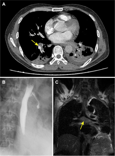
Figure 2 CT images of case 1 in September (A) and November (B). The enlarged mediastinal lymph node (arrow) decreased after antituberculosis therapy.
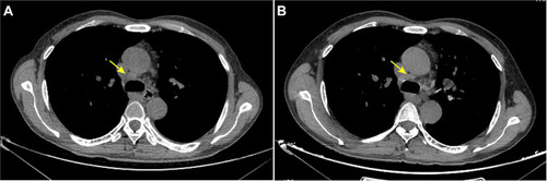
Figure 3 Imaging appearances in case 2. (A) The neck abscess (arrow) on axial MRI. (B) The right psoas abscess (arrow) on axial CT.
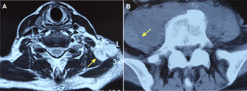
Table 1 Published Tuberculosis Cases Related to Ruxolitinib by the End of April 2020