Figures & data
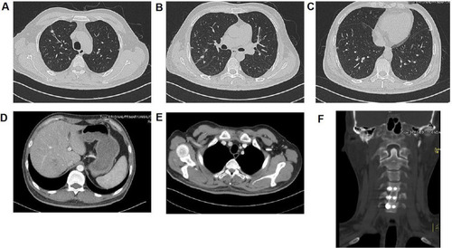
Figure 1 Enhanced computerized tomography (CT) of the implicated organs at admission. (A) Superior field of the lung; red arrow indicates multiple pulmonary nodules, partial liquefaction and necrosis; (B) Middle field of the lung; (C) Inferior field of the lung; (D) The right-posterior lobe of the liver; green arrow indicates abscess; (E) Right anterior superior chest wall; green arrow indicates abscess; (F) 5th cervical vertebra bone destruction with prevertebral abscesses; black arrow indicates bone destruction in the 5th cervical vertebra, and green arrow indicates prevertebral abscesses.
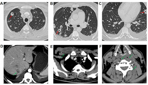
Table 1 Antimicrobial Susceptibility Tests and Interpretation Based on Blood, Sputum and Bronchoalveolar Lavage Fluid Isolates
Figure 2 Magnetic resonance imaging (MRI) of the cervical vertebra before operation. Green arrow indicates prevertebral abscess, and red arrow indicates bone destruction in the 5th cervical vertebra.
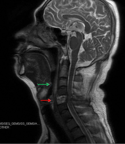
Figure 3 Postoperative H&E staining of the 5th cervical vertebra showing typical characteristics of chronic inflammation.
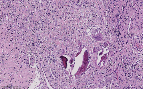
Figure 4 CT and MRI figures before second discharge. Focuses were significantly improved after treatment. (A) CT of the superior field of the lung; (B) CT of the middle field of the lung; (C) CT of the inferior field of the lung; (D) CT of the right-posterior lobe of the liver; (E) CT of the right anterior superior chest wall; (F) MRI of the 5th cervical vertebra.