
Figures & data
Figure 1 Chest X-ray. Chest diagnostic radiograph showed a slight increase and thickening of the texture of both lungs, with increased diffuse patch density. The boundary was fuzzy, and no obvious consolidation shadow was seen in the rest of the lungs.
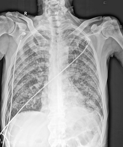
Figure 2 Partial Sanger sequencing peak map of the 16S rDNA PCR product of the isolate sequenced with the 1492R primer.
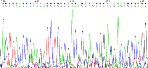
Table 1 Comparison of Case Reports of Mycobacterium colombiense Infections in Patients with and without HIV