Figures & data
Figure 1 Standard PA chest x-ray demonstrating widened superior mediastinum with homogenous opacity seen in the right upper lung zone.
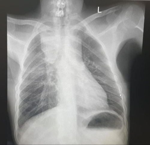
Figure 2 Microbiological summary timeline.
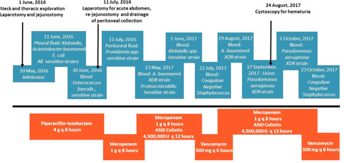
Table 1 Phenotypic Antimicrobial Susceptibility Pattern of the Strain PA179
Table 2 Summary of Genome Features of the Strain PA179
Figure 3 Circular Genome Representation of P. aeruginosa strain PA179. The DNAPlotter software from the Artemis Package was used standalone. Custom colors were assigned for the Visualization of tRNA and rRNA as well as for CDS, in the forward and reverse strands. The CDS in the forward strand is colored in Violet, CDS in Reverse is colored in blue, tRNA, and rRNA were colored in pink and orange small vertical lines, and the GC content and GC skew are colored in brown and violet, respectively.
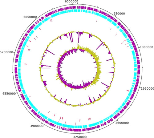
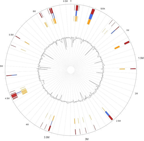
Figure 4 Genomic Islands in P. aeruginosa PA179.