Figures & data
Table 1 Primer Sequences and PCR Conditions
Table 2 Antibiotic Breakpoints
Table 3 Antibiotic Susceptibility and Typing of 65 Monoclonal Strains Isolated from 13 Patients
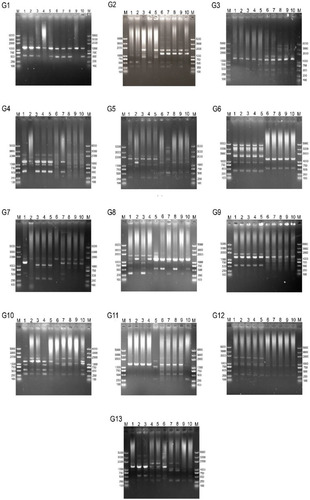
Figure 1 Electropherogram of RAPD-PCR typing of Helicobacter pylori strains from 13 patients.
Notes: M: 2K DNA ladder; G1–G13: sample/patient number; bands 1–5: monoclonal strains from a single patient amplified using primer 1247; and bands 6–10: monoclonal strains from a single patient amplified using primer 1283.