
Figures & data
Table 1 Gram-Negative Bacteria Demonstrating Imipenem Resistance Isolated from the Two Health Centers in North Gondar, Ethiopia
Figure 1 Percentage of culture positive and negative samples on MacConkey agar plates after 24 hours of growth.
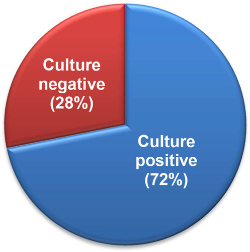
Figure 2 Diversity of Gram-negative imipenem-resistant bacteria identified by biochemical studies and 16S rRNA sequencing.
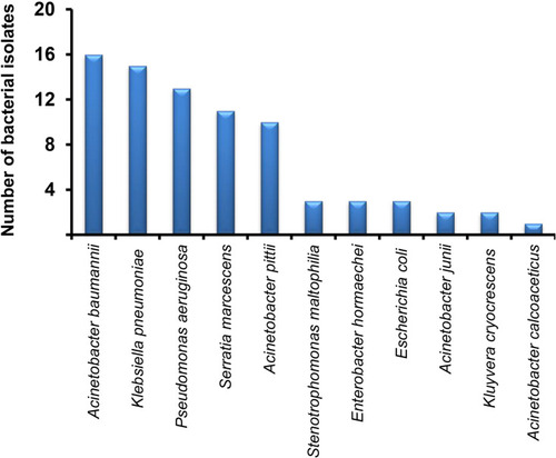
Figure 3 Comparison of the motility types of S. maltophilia isolates. The three different motility patterns was assayed using tryptone broth with 0.3% (w/v) agarose (swimming), nutrient broth with 0.5% (w/v) agar and 5 g/l glucose (swarming), and LB broth with 1% (w/v) granulated agar (twitching).
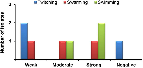
Table 2 Antimicrobial Resistance Pattern of Imipenem-Resistant Gram-Negative Bacteria Recovered from Sputum Samples
Table 3 Multidrug-Resistance Profiles of Imipenem-Resistant Gram-Negative Bacteria Recovered from Sputum Samples