Figures & data
Figure 1 (A) Timeline of case A progression, starting on the day of admission (days). After the administration of polymyxin B-immobilized fiber column-direct hemoperfusion (PMX-DHP) therapy twice on day 10, the patient’s dyspnea and respiratory rate improved on day 11. On day 13, the patient’s P/F ratio improved. (B) Timeline of case B progression, starting on the day of admission (days). After the administration of polymyxin B-immobilized fiber column-direct hemoperfusion (PMX-DHP) therapy on day 5, the patient’s dyspnea and respiratory rate improved on day 6. On day 8, the patient’s P/F ratio improved. Reference values: CRP is under 0.20 mg/dL, D-dimer is under 1.0 µg/mL.
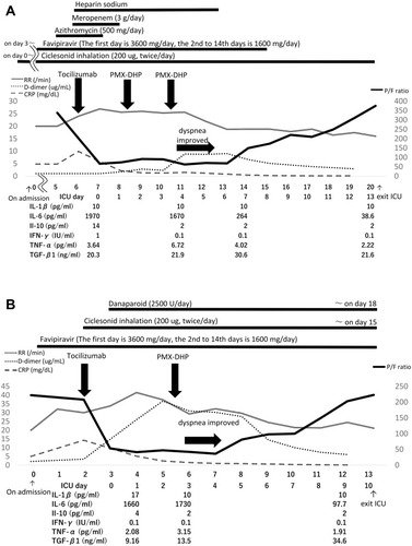
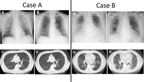
Figure 2 Serial chest X-ray and CT scans of cases (A and B). (A) CXR on day 7 (ICU day 0) shows worsening bilateral opacities. (B) CXR on day 88 shows normal lung parenchyma. (C) Chest CT scan on day 0 shows bilateral light GGOs. (D) Chest CT on day 95 shows disappearing bilateral GGOs, without the evidence of lung fibrosis. (E) CXR on day 3 (ICU day 0) shows the worsening of bilateral opacities. (F) CXR on day 13 shows improvement in aeration. (G) Chest CT scan on admission (day 0) shows light bilateral GGOs. (H) Chest CT scan on day 23 shows bilateral GGOs.