Figures & data
Figure 1 Procedure for synthesis of CM3a.1a: chromone. 2: maleimides. 3a: CM3a.
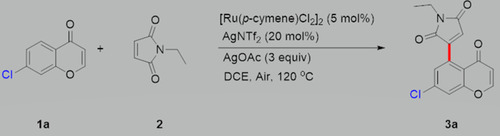
Figure 2 Growth assay for S. aureus strains treated with subinhibitory concentrations of CM3a. (A and B) Strains JP21 (A) and SA113 (B) were cultured with 4, 2, 1, 0.5, and 0.25 μg/mL CM3a or without CM3a for 24 h.
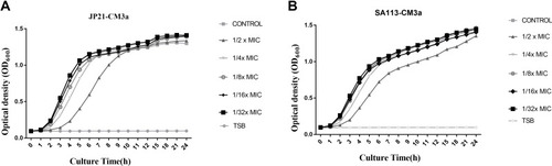
Figure 3 (A) Eradication of S. aureus (JP21, SA113, ZSA01, ZSA02, ZSA03, ZSA04, and ZSA05) biofilms by CM3a (16–128 μg/mL). (B) Eradication of S. aureus SA113 biofilms by vancomycin, telithromycin, and daptomycin. Each experiment was repeated 3 times, and data represent mean±standard deviation. **P<0.01, *P<0.05.
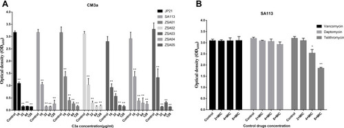
Figure 4 Biofilm formation after treatment with CM3a was observed by laser scanning confocal microscopy after the LIVE/DEAD assay. (A) Strain SA113 without treatment. (B) SA113 treated with dimethylsulfoxide (DMSO). (C) SA113 treated with CM3a at 2 times the MIC (2×MIC). (D) SA113 treated with CM3a at 4 times the MIC (4×MIC).
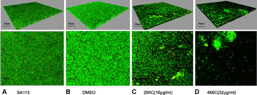
Figure 5 The survival rate of adherent cells in biofilms after treatment with CM3a (16–128 μg/mL). Each experiment was repeated 3 times, and data represent mean±standard deviation. **P<0.01.
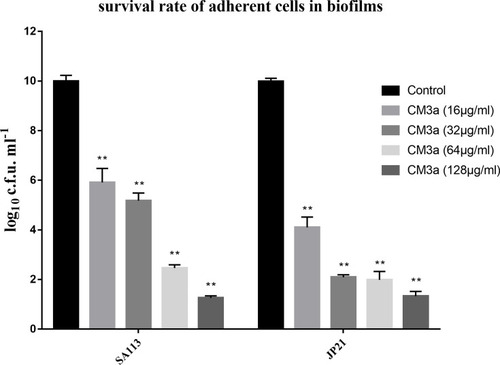
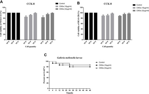
Figure 6 Toxicity of CM3a. (A and B) Viability of mouse alveolar epithelial cells MALE-12 (A) and human bronchial epithelial cell BEAS-2B (B) with or without CM3a treatment (16 and 32 μg/mL) as determined with the CCK-8 assay. (C) Survival of G. mellonella larvae following injection of CM3a (16 and 32 μg/mL) or PBS.