
Figures & data
Figure 1 (A) MRI of the lower extremities showed multiple clumpy abnormal signal lesions, suspected of hematomas or abscesses formation. (B) Abnormal signal lesions decreased than before. (C) Modified Kinyoun acid-fast stain (1% sulfuric acid as a decolorizing agent) revealed red purple branching hyphae in multiple directions (10*100). (D) A routine blood agar plate at 48 hours revealed growth of small and hard white colonies.
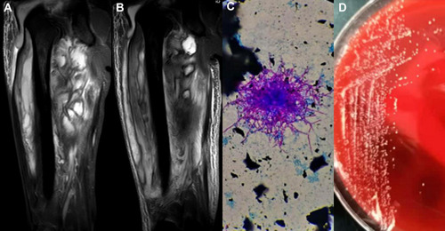
Table 1 Clinical Characteristics of 6 Patients with Primary Cutaneous Nocardiosis Due to N.farcinica Reported in Case Reports from 2006 to 2020
Table 2 Characteristics of the Patient from This Case Report