Figures & data
Figure 1 Neck CT findings. Mixed-density lesions on the left side of the neck (A). The masses were incised, and a drainage tube was inserted (B). Pus and necrotic materials (C). A large new exudative consolidation shadow was noted in the left upper lobe after 2 weeks of antibacterial treatment (D). Bronchofibroscopy revealed bronchial nodules in the left upper lobes that obstructed the lumen. Mucosal hyperplasia and hypertrophy were also noted (E).
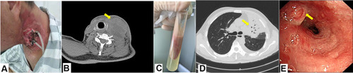
Figure 2 Talaromyces marneffei was isolated from the hilar nodes: Yellow colony with distinctive red diffusible pigment on Sabouraud’s dextrose slant at 25°C (A) and yellow colony on Sabouraud’s dextrose slant at 37°C (B). Lactophenol blue-stained culture of the ulcerating right supraclavicular subcutaneous mass showing that the conidiophores of this mold were smooth and had a size of 3 um, each of which had several phialides and produced smooth, spherical conidia in chains (C).
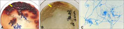
Figure 3 Computed tomography findings. Chest: The pulmonary lesions were markedly improved after 3 weeks of antifungal therapy (A). Neck: An abscess in the posterior pharyngeal wall, a fracture of the 4th cervical vertebra (B), and new mass on the right side of the neck (C) were found. The abscess disseminated to the lower right neck and sternum (D).
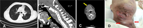
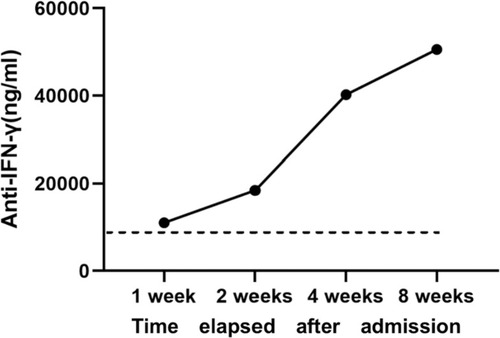
Figure 4 Neutralizing anti–interferon-γ autoantibody titers of the patient after admission. The dotted line indicates the level of the positive titer at each certain time point, peaking at 50,566 ng/mL at 8 weeks after admission.