Figures & data
Figure 1 Dermatologic examination. Appearance of vesicles with an erythematous base scattered on facial region (A–C), anterior and posterior trunk (D and E), upper extremity dextra (F and G) and sinistra (H and I), and dorsum manus (J).

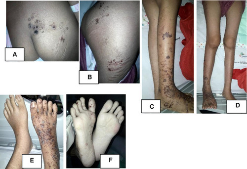
Figure 2 Dermatologic examination. Appearance of vesicles and some were confluent into bullae with erythematous base scattered on cruris dextra (A and D), lateral side of femur dextra (C), gluteus (B), and dorsum and plantar pedis dextra (E and F). Appeared striae on lateral side of right femur (C).
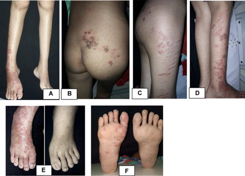
Figure 3 Tzanck smear. Appearance of MGC (
 ).
).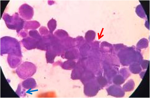
Figure 4 Follow up at 10th day. Showed hyperpigmented macules and patches on facial region (A–C), anterior (D) and posterior trunk (E), and upper extremity (F–I).
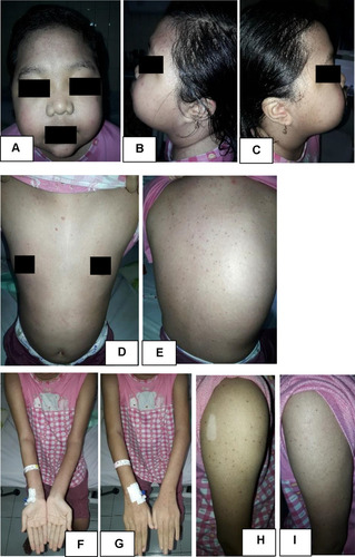
Figure 5 Follow up at 10th day. Showed hyperpigmented patches and and several sagging bullae, some burst into erosion covered by brownish crust at gluteus (A), lateral side of femur dextra (B), cruris dextra (C), and dorsum and plantar pedis region (D–F).