Figures & data
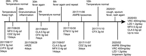
Figure 1 (A and B) shows lymph node enlargement in the hilus pulmonis zone; (B–E) shows that enlarged lymph nodes shrank gradually in the hilus pulmonis zone.
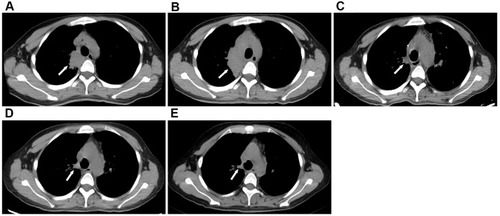
Figure 2 (A and B) pathological examination of H&E staining (H&E, 100×; H&E, 400×) from the cervical lymph nodes suggested granulomatous inflammation. Granulomas is composed of epithelioid cells in the lymph nodes. PAS(-).
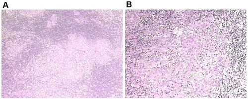
Figure 3 (A) Chest CT shows patchy infiltrates, banded and nodular shadows, pleural effusion. (A–C) Lesions in the lower lobe of the right lung and the pleural effusion absorbed gradually.
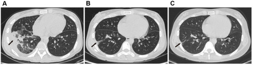
Figure 4 Treatment and manifestation timeline.
Abbreviations: CFX, cefoxitin; LVFX, levofloxacin; MFX, moxifloxacin; CDZ, cefodizime; CLA, clarithromycin; ETM, etimicin; H, isoniazid; R, rifampicin; Z, pyrazinamide; E, ethambutol; AMPB, amphotericin B liposomes; VRC, voriconazole; LZD, linezolid.