
Figures & data
Figure 1 Direct microscopic morphology of skin tissue and CSF. (A) Direct wet film microscopy of skin tissue, 400×. (B) Gram stain of skin tissue, 1000×. (C) GMS of skin tissue, 1000×. (D) GMS of CSF after centrifugation, 1000×.
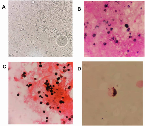
Figure 2 Colonies and microscopic morphology of cultured skin tissue and CSF specimens. (A) Colonies on CHROMagar Candida medium from skin tissue cultures. (B) Wet film microscopy of skin tissue cultures, 40×. (C) Gram stain of skin tissue cultures, 1000×. (D) Colonies on blood agar from CSF cultures.
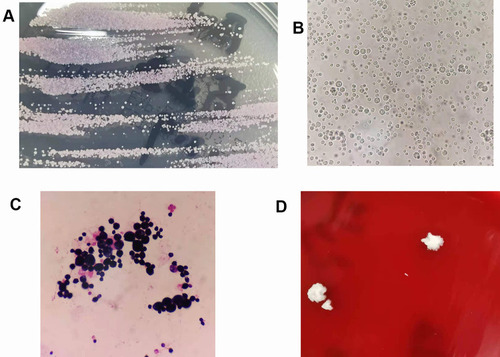
Table 1 Reports of Meningitis Due to Prototheca spp. Up to April 2021