
Figures & data
Table 1 Sequences of the Primers and the Size of Amplicon in Base Pair (Bp) Used in Multiplex PCR for Detection of UPEC Strains Phylogroups and PAIs
Table 2 Comparison of the Phylogenetic Groups, PAIs, Antibiotic Resistance Pattern and Biofilm Formation of UPEC Isolated from CAUTI and Com-UTI
Figure 1 Detection of UPEC phylogroups using multiplex PCRCitation11 targeting the genes chuA (279bp) and yjaA (211bp) and the DNA fragment TspE4.C2 (154bp). Lane M: 100 bp DNA Ladder. Lane 1: Distilled water as negative control, Lane 2: phylogroup A, Lane 3: phylogroup B1, Lane 4,5: phylogroup A, Lane 6: phylogroup B1, Lane 7: phylogroup B2, Lane 8: phylogroup A, Lane 9: phylogroup A, Lane 10: phylogroup D, Lane 11: phylogroup A, Lane 12: phylogroup B2 and Lane 13: phylogroup A.
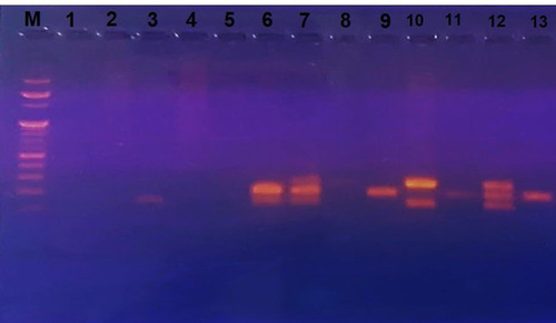
Figure 2 Detection of UPEC PAIs using multiplex PCR A to detect PAI III536 (200 bp), PAI IV536 (300 bp) and PAI IICFT073 (400 bp). Lane M: 100 pb DNA ladder. Lane 1: UPEC strains with PAI IICFT073, Lane 2: UPEC strains with no PAI, Lane 3: UPEC strains with PAI IV536 and PAI IICFT073, Lane 4: UPEC strains with no PAI and Lane 5: UPEC strains with PAI IV536.
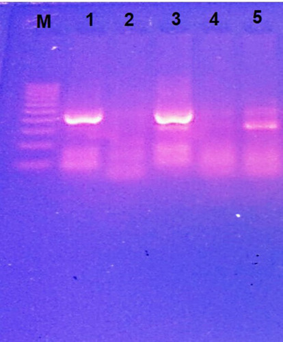
Figure 3 (A and B) Detection of UPEC PAIs using multiplex PCR B to detect 5 PAIs; PAI IJ96 (400 bp), PAI ICFT073 (930 bp), PAI II536 (1000 bp), PAI I536 (1800 bp) and PAI IIJ96 (2300 bp). (A); Lane M: 100 pb DNA ladder. Lanes 1–5: UPEC strains with PAI ICFT073. (B); Lane M: 100 pb DNA ladder. Lanes 1–6,8: UPEC strains with no PAI and Lanes 7,9 UPEC strains with PAI IJ96.
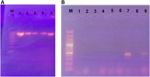
Table 3 Antibiotic Resistance Pattern and Biofilm Formation of Different UPEC Phylogroups Isolated from CAUTI and Com-UTI
Table 4 Prevalence of PAIs in Different UPEC Phylogroups Isolated from CAUTI and Com-UTI