Figures & data
Table 1 Patients’ Clinical Characteristics
Table 2 CT Characteristics of Solitary and Multiple Nodules
Figure 1 A 24-year-old woman with HIV infection and pulmonary cryptococcosis. She has headache and fever for ten days, and cryptococcal meningitis is confirmed. (A and B) Axial CT images show multiple clustered nodules with different sizes located in the subpleural zone of right lower lobe. (B) Cavities are detected in the bigger nodules.
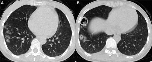
Figure 2 A 24-year-old man with HIV infection and pulmonary cryptococcosis. He has headache and fever for a week, and cryptococcal meningitis is confirmed. (A and B) Axial CT images show multiple scattered nodules with different sizes in the right lower lobe, and cavities are detected in the bigger nodules.
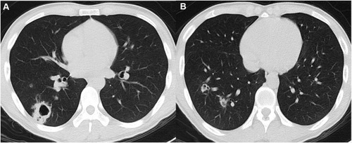
Figure 3 Patients with pulmonary cryptococcosis presented as solitary solid nodules. (A) Axial CT image in a 48-year-old asymptomatic man shows a round and well-defined nodule with small bronchi located in the right upper lobe. (B) Coronal CT image in a 54-year-old asymptomatic man shows a round and well-defined nodule with vacuole located in the left upper lobe. (C) Coronal CT image in a 50-year-old asymptomatic man shows an ill-defined nodule with a satellite lesion (red arrow) located in the right upper lobe. (D) Axial CT image in a 49-year-old asymptomatic woman shows an oval nodule with halo sign located in the left upper lobe.
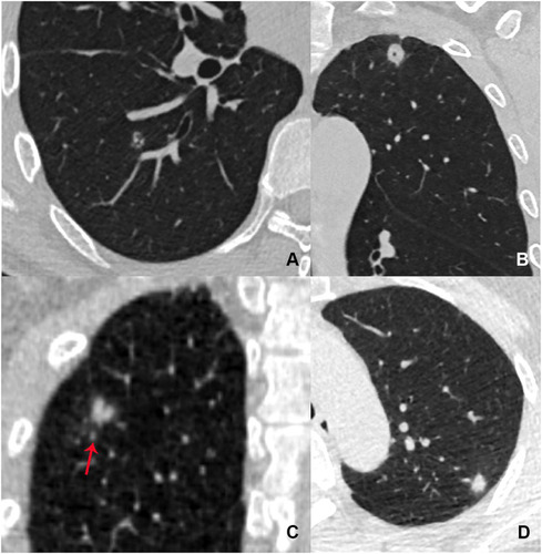
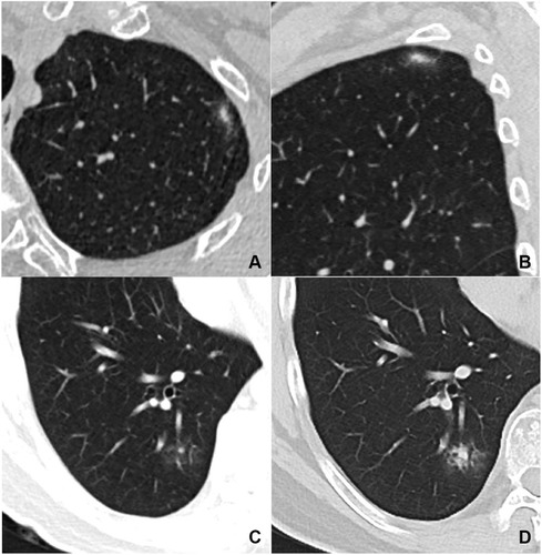
Figure 4 Patients with pulmonary cryptococcosis presented as solitary subsolid nodules. (A and B) Axial and sagittal CT images in a 60-year-old asymptomatic woman show an oval and ill-defined mixed GGN with strip shaped solid component located in the left upper lobe. (C and D) Axial CT images in a 50-year-old asymptomatic man show an ill-defined mixed GGN located in the right lower lobe. (D) The solid component in this nodule significantly increases after one month.