Figures & data
Figure 1 Growth curve and the biofilm formation of six VV isolates. (A) growth curve; (B) biofilm formation.
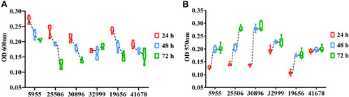
Figure 2 Schematic diagram of one genomic island in five VV isolates. The arrows represent the positions and direction of the elements.
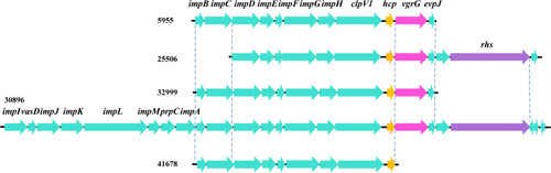
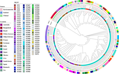
Figure 3 Core-genome-based phylogenetic tree of 163 VV isolates, including 6 isolates from this study and 157 strains downloaded from NCBI genome database. STs of the isolates is labelled in the outer ring. The source of all isolates is presented in the middle ring. The location of the isolates is colored in the inner ring. The number of isolates in the present study was colored in red.
Abbreviations: NA, not available; NEW, novel STs.