Figures & data
Figure 1 The epidemiological distribution of cystic and vesicular cysticercosis in China.
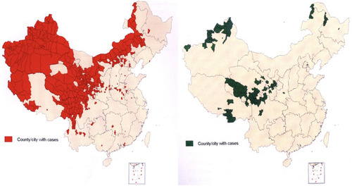
Figure 2 The pathological changes in cystic echinococcosis: When the course of echinococcosis was long, the external capsule of the Echinococcus often became thick and rough with calcium salt deposits or even completely calcified. The Echinococcus had mostly decayed and necrosed, with purulent cystic fluid. When the Echinococcus gradually degenerated with the absorption of the cystic fluid, the inner cyst wall folded and shrank, and the necrosis dissolved with caseous changes.
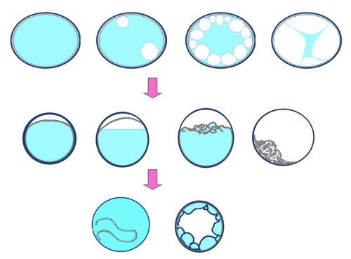
Figure 3 Cystic echinococcosis—the simple cystic type.
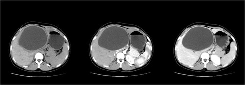
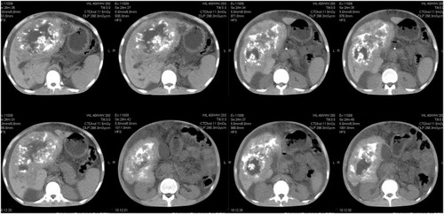
Figure 4 Cystic echinococcosis—the multi-daughter cystic type.
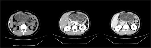
Figure 5 Cystic echinococcosis—the inner capsule detached type.
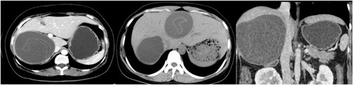
Figure 6 Cystic echinococcosis—postoperative recurrence, rupture, combined with infection.
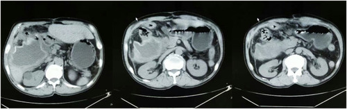
Figure 7 Alveolar echinococcosis—the annual ring sign.
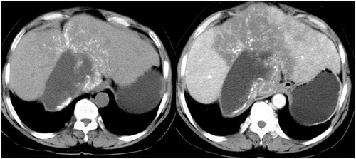
Figure 8 Alveolar echinococcosis—the CT and MRI manifestations. (A) CT imaging: A marginal, hypodense infiltration zone is observed, coexisting with both calcified deposits with irregular masses at the inner margin and a cavity. (B and C) MRI: The infiltration zone manifests as a hypodense irregular mass on T1- and T2-weighted images and the inner edge of the fibrous zone manifests as slightly hyperdense and locally invaginated to form a peninsula sign, with internal necrosis and liquefaction forming a fluid retention cavity.
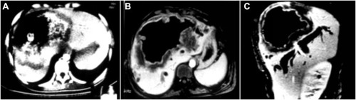
Figure 9 Alveolar echinococcosis—the vesicle sign and peninsula sign.
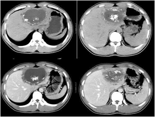
Figure 10 Alveolar echinococcosis—with multiple typical signs.