Figures & data
Table 1 Distribution of rpoB Mutations and MICs of Rifampin
Table 2 Logistic Regression Multivariate Model Results Between rpoB Mutations and RIF Resistance
Table 3 Summary for Interaction Changes of Mutations Based on Structural Analysis
Figure 1 The 2D diagram showing the interactions between rifampin and wild-type RpoB (A), mutant S441L (B) or double mutant D435E & S441L (C). The rifampin (RIF) molecule is shown in the middle with a display style of ball and stick. The colored balls around RIF molecule indicate the residues involved in the direct interactions between RpoB and RIF. The green, magenta and red dash lines connecting RIF and corresponding residues indicate intermolecular hydrogen bonds, hydrophobic interactions and steric hindrance, respectively. Residues involved in van der Waals interactions are represented by light green balls without any dash line linked to RIF.
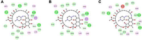
Figure 2 Interactions between rifampin and Residue 445 of wild-type/mutant RpoB. The detailed structure for rifampin (RIF) and Residue 445 of wild-type RpoB H445 (A), mutant H445R (B), mutant H445L (C), mutant H445Y (D), mutant H445C (E), mutant H445D (F), mutant H445P (G), mutant H445N (H) or mutant H445Q (I). Residue 445 of RpoB is shown in the left with a display style of Corey-Pauling-Koltun (CPK). The RIF molecule is shown in the right with a display style of stick. The intermolecular hydrogen bonds are indicated by green dashed lines.
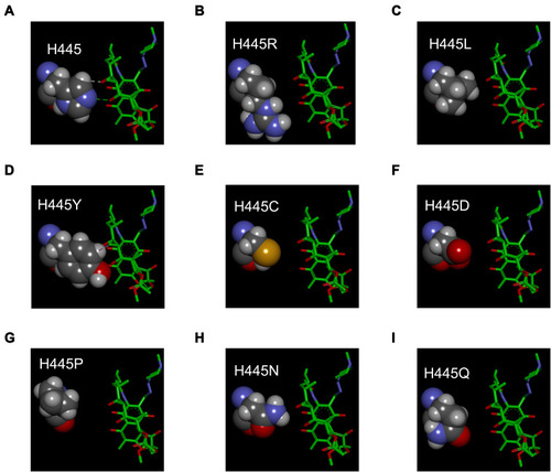
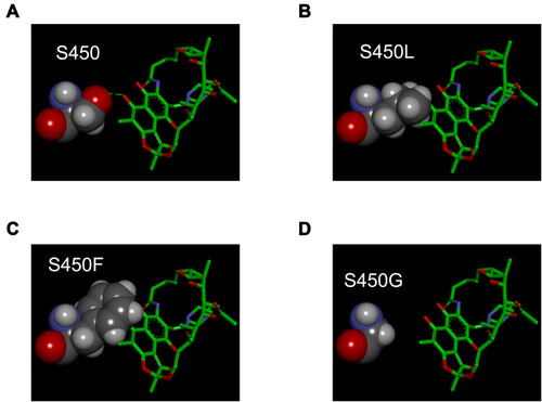
Figure 3 Interactions between rifampin and Residue 450 of wild-type/mutant RpoB. The detailed structure for rifampin (RIF) and Residue 450 of wild-type RpoB S450 (A), mutant S450L (B), mutant S450F (C) or mutant S450G (D). Residue 450 of RpoB is shown in the left with a display style of Corey-Pauling-Koltun (CPK). The RIF molecule is shown in the right with a display style of stick. The intermolecular hydrogen bonds are indicated by green dashed lines.