
Figures & data
Figure 1 Vacuum-assisted closure (VAC) in the treatment of deep neck infection. Simultaneous VAC: (A) The abscess cavity was exposed, and necrotic tissues were removed. (B) The foam material was then placed into the infected area, and the transparent film completely covered the wound to ensure sealing. In addition, the VAC device was connected. Staged VAC: (C) Infection wound failed to heal after conventional drainage and repeated debridement. (D) The VAC device was then placed into the infected area to facilitate wound healing. White arrows in (B and D) indicate the foam material and the transparent film, respectively.
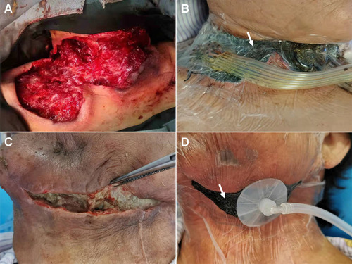
Table 1 General Information
Table 2 Characteristics of Infection
Table 3 Evaluation of Efficacy