Figures & data
Figure 1 LAM TB (upper) and cryptococcal antigen test (lower) results.
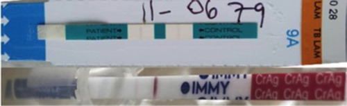
Figure 2 Anteroposterior chest radiography demonstrated diffuse reticulonodular opacities (nodular> reticular) involving the bilateral hemithorax with background ground glass haziness.
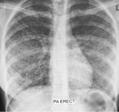
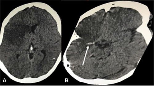
Figure 3 Unenhanced computed tomography of brain, (A) axial image at the level of anterior horns of the lateral ventricles demonstrating a large geographic area of hypodensity involving the right frontopareitotemporal cortex. (B) axial image at the level of circle of Wills demonstrating hyperdense middle cerebral artery sign (white arrow) of the M1 segment of the right middle cerebral artery.