Figures & data
Figure 1 (A and B) Redness and swelling of the forearm. (C and D) MRI revealed marked forearm muscle swelling, heterogeneous signal intensity and shadowing suggestive of gas.
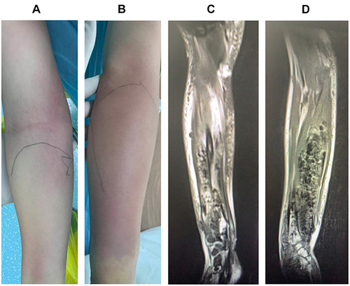
Table 1 List of Primers Used for Detection of Hypervirulent K. pneumoniae Virulence Genes
Figure 2 (A and B) Pus and gas were observed within the forearm musculature and intermuscular space during surgery. (C and D) Partial muscular necrosis and infection of the interosseous membrane and periosteum was observed.
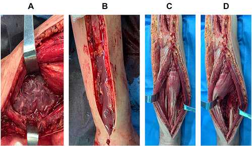
Figure 3 The string test was performed using an inoculation loop to evaluate for hypermucoviscosity. (A) Formation of mucoviscous strings >5 mm in length was defined as a positive result. (B and C) Agarose gel electrophoresis revealed that the strain carried rmpA, rmpA2, iucA, iroB, and PEG-344 virulence genes and belonged to the K1 capsular serotype.
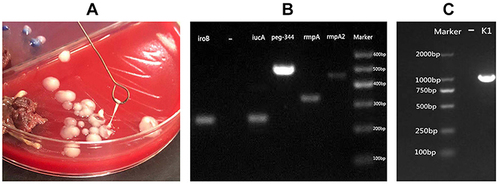
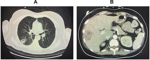
Figure 4 (A) Chest CT showing a lung abscess. (B) Abdominal CT showing a multilocular liver abscess.