
Figures & data
Figure 1 Clinical and pathology examination of skin lesions presented by the patient. (A) Maculopapular rash on the lower limbs with Biett collarette; (B) erythema nodosum on the right forearm; (C) granulomatous dermatitis with a lichenoid infiltrate; (D) septal panniculitis.
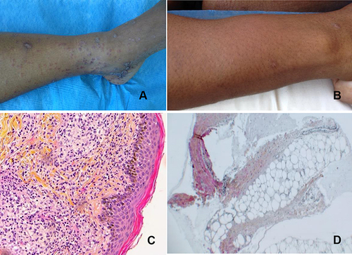
Table 1 Case Reports of Erythema Nodosum Associated with Syphilis