Figures & data
Figure 1 Swelling and purulent exudation from the wrist (A) to the left forearm (B); beaded proliferative nodules from the dorsum of the right hand (C) to the right forearm (D).
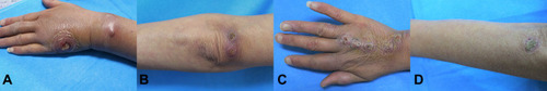
Figure 2 Scattered nodules on both knees (A) and the left lower limb (B).
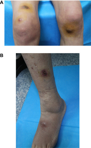
Figure 3 The histopathological results showed superficial and deep granulomatous dermatitis with suppuration, multinucleated giant cells (A); negative periodic acid Schiff (PAS) staining (B); acid-fast bacilli were seen on skin biopsy stained with Ziehl–Neelsen stain (C).
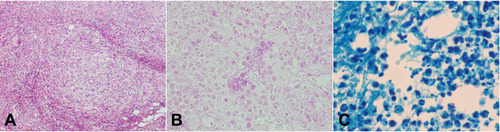
Figure 4 White, smooth colonies was observed in potato dextrose agar (PDA) medium after 1 week.
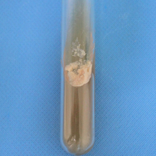
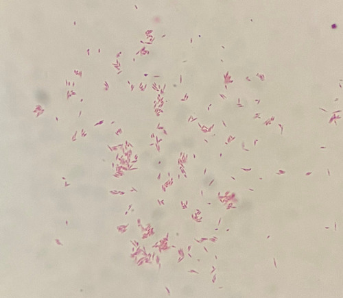
Figure 5 Acid-fast staining of the bacterial suspension demonstrated acid-fast bacilli.