Figures & data
Figure 1 (A) Optical density (OD) of S. aureus with different conc. of L. inermis total extract (MIC = 0.0725 mg/mL). (B) OD of S. aureus with different conc of L. plantarum supernatant (MIC=13mg/mL. (C) OD of S. aureus with different conc. of L. plantarum supernatant and L. inermis extract (1:1), (MIC = 2 mg/mL).
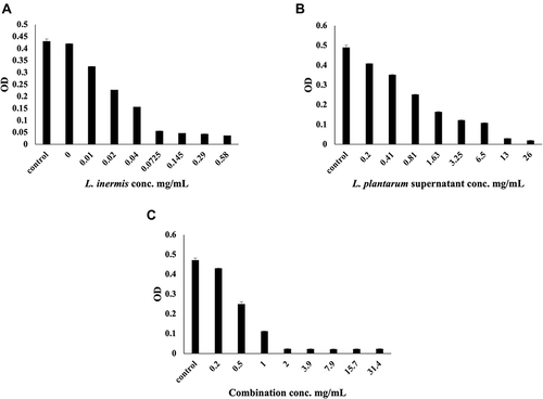
Table 1 IL-6, TNF-α Levels and WBCs Count of All Groups
Table 2 Parameters of Histologic Assessment of Wound
Figure 2 Histopathological examination of non infected wound before and after treatment with L. plantarum and or L. inermis. (A and B) Uninfected group. Sections showed a shallow ulcerative lesion with minimal epidermal and dermal florid inflammatory reaction. All other structures of the skin keep a normal morpho-histological appearance. (C and D) Lactiplantibacillus cell group. The skin specimens revealed normal morpho-histological structures; however mild superficial contamination was seen, but without any significant pathologic reaction. (E and F) that replaced large parts of the epidermis was seen. The ulcerated tissue is replaced by suppurative inflammatory exudate with a predominance of neutrophils and secondary saprophytic infection. (G and H) Lawsonia Group. Examined sections revealed a mild superficial exfoliative reaction with secondary contamination. (I and J) Combination group. Although clear ulcerative lesions were not seen, a peculiar exudative epidermal reaction was seen. Such exudate was seen covering the epidermis and contained a moderate number of neutrophils and the saprophytic contaminant. An epidermal exfoliative reaction was also seen (H&E X400, zoomed section X30).
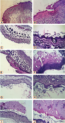
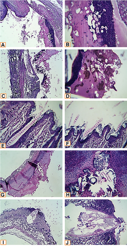
Figure 3 Histopathological examination of infected wound before and after treatment with L. plantarum and or L. inermis. (A and B) Infected group. Sections showed deep ulcerative lesions with complete exposure of the epidermis and extending to the dermis. An extensive inflammatory reaction with secondary infection and predominance of neutrophilic infiltration were seen. (C and D) Lactiplantibacillus cell group infected with S. aureus. A wide detached ulcer was seen overlying denuded dermal tissue. The ulcerated materials were mostly formed from suppurative exudate rich in neutrophils and contained contaminated saprophytes and amorphous brownish materials (bloody-like). (E and F) Lactiplantibacillus supernatant group infected with S. aureus. The skin structures were completely normal. An elongated septated eosinophilic contaminant of unknown nature was seen overlying the epidermis and superficially invaded it. (G and H) Lawsonia group infected with S. aureus. A huge detached ulcerative tissue was seen. It was formed from suppurative exudate rich in neutrophils, bacterial colonies, dead necrotic tissue, and saprophytic structures. (I and J) Combination group infected with S. aureus. A characteristic narrow, deep ulcerative lesion was seen. It extended deep to the hypodermis. The contents of the ulcer were funneled shape hyalinized membranous structures entangling necrotic debris. Neutrophils extensively infiltrated the adjacent skin tissue; other parts of the skin in the vicinity of the ulcer showed a florid inflammatory reaction, enclosing different types of leucocytes (H&E X400, zoomed section X30).