Figures & data
Figure 1 The clinical course of our patient. The clinical course includes the therapy for NTM-PD, the smear culture and NTM strain, clinical disease activity index (CDAI) of rheumatoid arthritis, the changes in the levels of Krebs von den Lungen-6 (KL-6, blue line) and C-reactive protein (CRP, Orange line), and longitudinal chest imaging findings (A) at referral, (B) at pyrothorax, (C) at the diagnosis of rheumatoid arthritis (RA), (D) at abatacept introduction, and (E) at 1 year after initiating abatacept therapy.
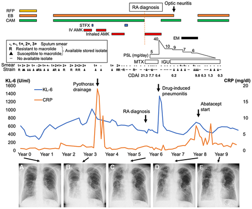
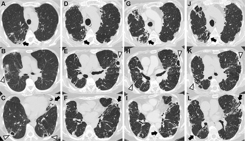
Figure 2 Chest computed tomography (CT) findings during the clinical course. (A–C) At referral, multiple cavitary lesions (white arrowheads) are in both lungs with bronchiectasis (black arrows) in lingula. (D–F) At the diagnosis of rheumatoid arthritis (RA), cavitary lesions and bronchiectasis are worsening. (G–I) At the introduction of abatacept, no change has occurred in the cavitary lesions in the left lung or bronchiectasis in bilateral fields. (J–L) One year after the introduction of abatacept, cavitary lesions partially improved, whereas a new small cavity has developed in the right lung.