Figures & data
Table 1 The Primers and Their Sequences for Integron Screening
Table 2 The Distribution of Class 1 and 2 Integrons and Gene Cassettes in 150 Strains of Clinically Isolated Proteus mirabilis
Figure 1 The functional class 2 integrons. (A) General structure of the functional class 2 integrons: Arrows indicate the coding sequences with the gene name above, triangles and circles are attC and attI recombination sites, respectively. The attI2 region and gene cassette array are indicated. Dotted vertical bars separate each gene cassette. (B) Nucleotide sequence of the attI2 region. The −35 and −10 elements of the Pc2 promoters are written in bold uppercase, and their names are indicated. The transcriptional mapped for PintI2 is indicated by a Broken Arrow and bold uppercase. The position of several nucleotides is numbered (italics).
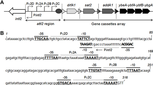
Table 3 Comparison of Antimicrobial Resistance Rates of 150 Strains of Proteus mirabilis with Major Resistance Genes in Variable Regions (%)
Table 4 Three Functional Class 2 Integron-Related Drug Resistance Genes and Phenotypes
Figure 2 The mutation of the internal stop codons of the functional class 2 integrons: the stop codons in these intI2 genes were mutated from TAA to the glutamine codon CAA. The position of several nucleotides is numbered (italics).
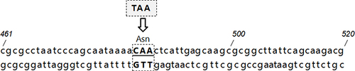
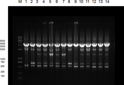
Figure 3 ERIC-PCR typing map of some class 2 integron-positive strains:M represents DNA Marker. Lane 1 is a functional class 2 integron-positive quality control strain; lanes 2∼4 are functional class 2 integron-positive strains; lanes 5∼14 are class 2 integron-positive strains; lanes 3∼6 and 10∼14 are type A strains, lanes 2 and 8 are of type B, lane 7 is the type C and lane 9 is a type D strain.