Figures & data
Table 1 Demographic and Epidemiological Data of Human Sporotrichosis Cases (n = 4969) Diagnosed in Jilin Province, China During 1990–2019
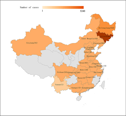
Figure 1 Distribution of sporotrichosis in Jilin Province, China, during1990–2019. The data from the Second Hospital of Jilin University diagnosed sporotrichosis, total 4969 cases.
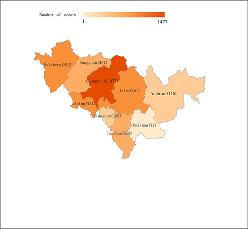
Figure 2 Changes in the visits of patients with sporotrichosis in Jilin Province, China, during 1990–2019.
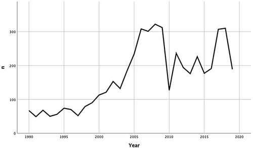
Figure 3 Clinical manifestations of sporotrichosis in patients with chronic cutaneous and subcutaneous lesions in Jilin Province, China (A) Fixed lesions: solitary or satellite erythematosus and papular lesions. (B) Lymphocutaneous lesions: ulcerative/papular/tumor-like lesions distributed along the lymphatic vessels. (C) Disseminated lesions: ulcerative/granulomatous/crusty lesion with multiple cutaneous lesions.
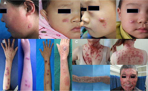
Table 2 Correlation of the Clinical Type with Age, Location, and Disease Course
Table 3 Bivariable and Multivariable Logistic Regression of Factors Influencing Sporotrichosis Types
Figure 4 Mycological culture of sporotrichosis. (A) Colony morphology of isolated Sporothrix schenckii grown on potato dextrose agar at 25°C following incubation for 2 weeks. (B) The isolated strain was identified using microscopy with lactophenol cotton blue staining.
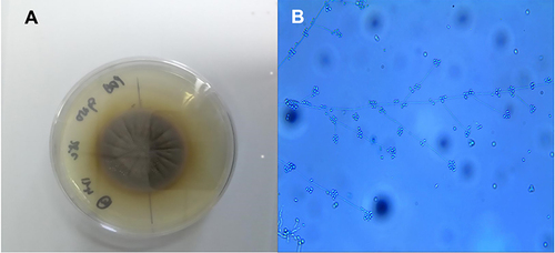
Table 4 The Distribution of Sporotrichosis in China
Figure 5 Flowchart of the literature search.
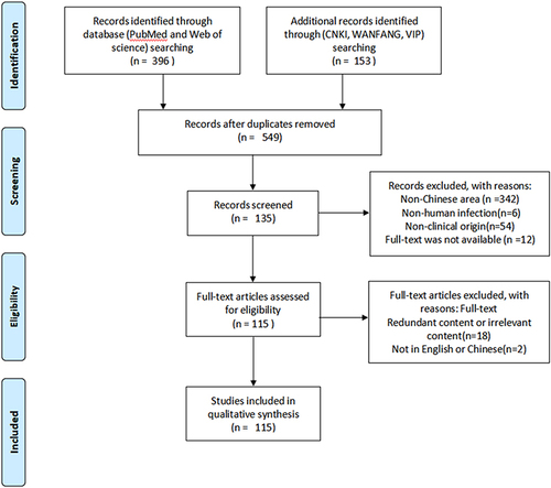
Figure 6 Distribution of sporotrichosis in China (2010–2019). The data are from the literature search depicted in Supplementary Table 1, including 17 studies in English and 98 in Chinese.