Figures & data
Table 1 Baseline Characteristics of Patients with SARS-CoV-2 Infection After Vaccination
Table 2 Demographic and Clinical Characteristics of SARS-CoV-2 Patients with Fully and Partly Vaccinated
Figure 1 The time interval between vaccination and infection in patients with SARS-CoV-2 infection. Patient number color in red was symptomatic infection and number in black was asymptomatic infection. * Patient with partly vaccinated.
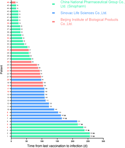
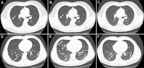
Figure 2 Representative chest CT images of two confirmed COVID-19 patients. A 34-years-old male with mild COVID-19. (A) Chest CT images showing right lung small patchy shadows on admission. (B) The chest imaging alleviated on the third day after admission. (C) Chest CT images complete remission 7 days after admission. A 30-years-old male with moderate COVID-19. (D) Chest CT images showing bilateral ground-glass opacity and patchy shadows on admission. (E) The chest imaging was aggravated, accompanied by vascular shadow and bronchiectasis in the focus on the third day after admission. (F) Chest CT images showing bilateral ground-glass opacity and patchy shadows was improved and the inflammatory exudation was absorbed 20 days after admission. White arrows showed the imaging lesion on chest CT.