
Figures & data
Figure 1 Enhanced computed tomography of the neck. Black arrow indicates a right peritonsillar abscess. Black arrowhead shows a retropharyngeal abscess.
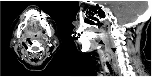
Figure 2 Enhanced computed tomography of the neck. Black arrow indicates right internal jugular vein thrombosis.
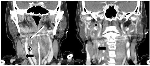
Figure 3 Clinical course of the present case.
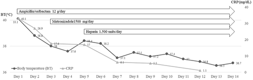
Table 1 Primers Used for 16S rRNA SequencingCitation8
Table 2 Antimicrobial Susceptibility of Dialister pneumosintes Isolated in the Present Case. Breakpoint Was Measured Based on Clinical and Laboratory Standards Institute M100-S26
Figure 4 Colony morphology of the present strain on Brucella HK agar plate.
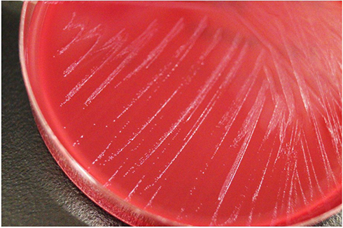
Figure 5 16S rRNA sequence of the isolated strain.

Figure 6 Hit taxon and strain name of the isolated strain.
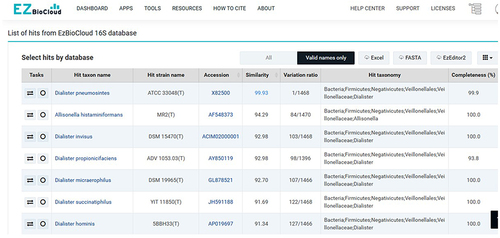
Table 3 Reported Cases of Dialister pneumosintes-Associated Bacteremia, Including the Present Case