Figures & data
Figure 1 (A and B) MRI of lumbar spine displayed partial cortical bone destruction and formation of abscesses at L4. (C) A chest CT scan demonstrated small round nodules with uneven density of upper right lung. The red arrow represents lesion.
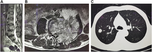
Figure 2 (A) Hematoxylin-eosin (HE) staining of the tissue sample from punch biopsy on L4 spine; the red arrow represents Cryptococcus. (B) Periodic acid–Schiff staining of the tissue sample from surgical biopsy on L4 spine.
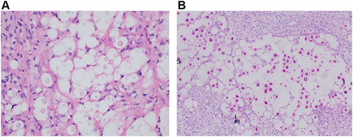
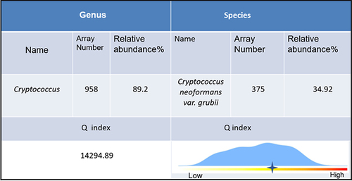
Figure 3 Next-generation sequencing (NGS) data.