
Figures & data
Figure 1 Chest X-ray (A) and computed tomography (CT) (B) findings of patient 1. Arrows indicate the abnormal small nodules, infiltrate shadows, and bronchiectasis.
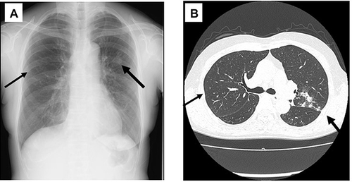
Figure 2 Colonies and stained fungal bodies of Exophiala dermatitidis from the bronchial lavage fluid of patient 1. Small colonies (A) and the filamentous and yeast-like fungi (B) were found by Gram staining at 24 hours later. The annelloconidia are clearly seen with lactophenol cotton-blue staining after 48 hours incubation of the small colonies at 24 hours with human leucocytes as the stimulator (C). Huge, dark colonies (D) and yeast-like form fungi (E) were found at 72 hours later, The colonies at 72 hours show high viscosity ((F), arrows).
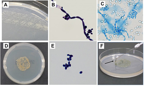
Figure 3 Matrix-assisted laser desorption ionization time-of-flight (MALDI-TOF) mass spectrometry (MS) analysis of the isolated black fungus. The fungus matches with a high score value (>2.0) as Exophiala dermatitidis.
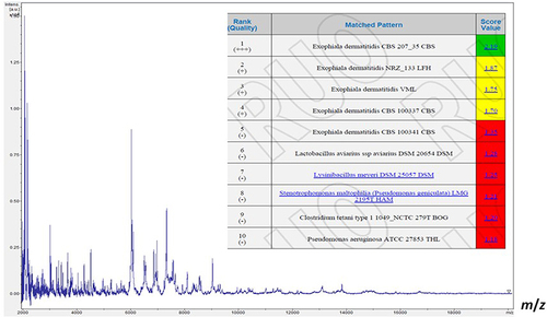
Figure 4 Chest and facial x-ray and computed tomography (CT) findings of patient 2. Chest x-ray (A) and CT (B) show only small nodules and slight bronchiectasis in both middle-lower lung fields, respectively (arrows). Facial x-ray (C) and CT (D) suggested the right sinusitis. Arrows indicate the suggested lesions of the right paranasal sinus.
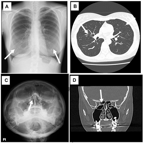
Table 1 Minimum Inhibitory Concentrations (MICs) of Various Antifungal Agents for the Exophiala dermatitidis Isolated from the Two Patients